Human Bone Anatomy Chart ~ Vintage Poster The Human Skeleton Muscles Of The Human Body Anatomy Chart 80 S Ebay. Check out pictures and diagram related to bones, organs, senses, muscles and much more. The bones of the axial skeleton act as a hard shell to protect the internal organs—such as the brain and the heart—from damage caused by external forces. This diagram depicts skeletal images 744×1314 with parts and labels. This article looks at the anatomy of the back, including bones, muscles, and nerves. The anatomy of the femur can be divided into proximal, central, distal, and posterior parts.
This diagram depicts human skeleton with parts and labels. Muscle anatomy reference charts free pdf download kenhub from thumbor.kenhub.com explore the world of human anatomy for kids with these colorful the first lesson of the anatomy for kids free printable file, starts with an introduction to the bone system chart. Bone zygomatic bone maxilla mandible nasal bones perpendicular plate of ethmoid nasal conchae note the nasal bones only make up a small portion of the bridge of the nose, most of the external nose is cartilage. Posted on may 28, 2014 by admin. Learn more about human anatomy with these free resources.
Vintage 3d Human Body Chart Skeletal System Human Skeleton Anatomy Thirdshiftvintage Com from cdn.shopify.com Flat vector medical illustration isolated. The femur and/or hip may fracture secondary to trauma, so understanding the femur bone anatomy is important. (spanish version) the human muscular system anatomy chart is a gorgeous yet complete guide to the human muscular system, displaying a human figure from front and back. All anatomy charts are available in 19.7 x 26.6 in (50 x 67 cm) unless otherwise stated. Explore the anatomy systems of the human body! Altogether, the skeleton makes up about 20 percent of a person's body weight. There are numerous types and combinations of these worksheets, and they can be found in virtually every medical classroom, no matter size or age the students. The human skeletal system consists of all of the bones, cartilage, tendons, and ligaments in the body.
Human anatomy bones worksheets are a fun and useful way to simply help students understand the anatomy of these body.
The femur and/or hip may fracture secondary to trauma, so understanding the femur bone anatomy is important. It also covers some common conditions and injuries that can affect the back. Posted on january 9, 2021 by kids. Anatomy chart of human bones for medicine and health care themes design. Posted in diagrams | tagged all bones, human skeleton, skelet, skeleton human eye featured. There are numerous types and combinations of these worksheets, and they can be found in virtually every medical classroom, no matter size or age the students. This diagram depicts human skeleton with parts and labels. Posted on may 28, 2014 by admin. 12 photos of the finger diagram bones. Anatomical wall charts and posters from 3b scientific® are ideal for teaching human anatomy, patient education and medical studies! A table on the poster also lists the main organs and their function as part of the digestive system. The bones of the appendicular skeleton provide support and flexibility at the joints and anchor the muscles that move the limbs. Altogether, the skeleton makes up about 20 percent of a person's body weight.
(spanish version) the human muscular system anatomy chart is a gorgeous yet complete guide to the human muscular system, displaying a human figure from front and back. Anatomy chart of human bones for medicine and health care themes design. Clearly labelled through out, but without cluttering the anatomy itself, this is an ideal anatomy poster for classrooms to treatment rooms. This colorful anatomical chart illustrates the human gastrointestinal system. The femur and/or hip may fracture secondary to trauma, so understanding the femur bone anatomy is important.
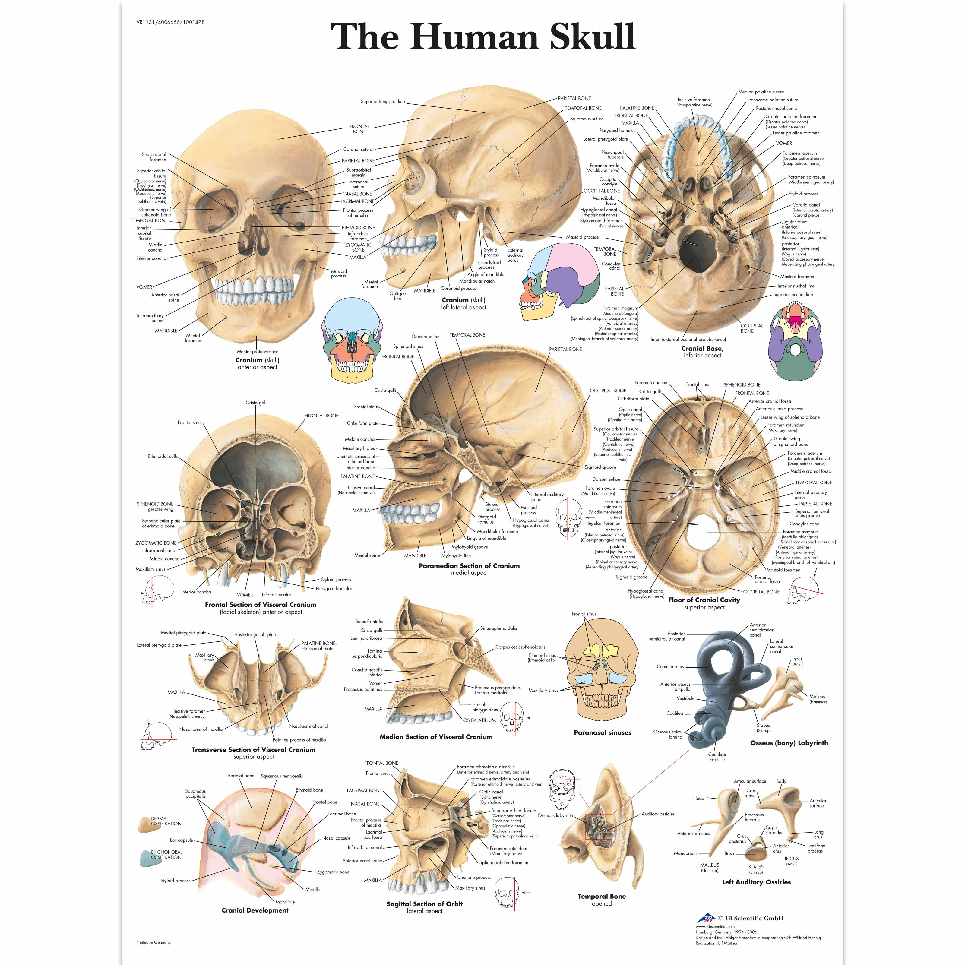
Human Skull Chart 4006656 Vr1131uu Skeletal System 3b Scientific from www.3bscientific.com The femur and/or hip may fracture secondary to trauma, so understanding the femur bone anatomy is important. (spanish version) the human muscular system anatomy chart is a gorgeous yet complete guide to the human muscular system, displaying a human figure from front and back. Explore the anatomy systems of the human body! The human heart is found just to the left of the breastbone and is about the size of a fist. 10 human anatomy bones worksheets. This framework consists of many individual bones and cartilages. Anatomy chart of human bones for medicine design. It also covers some common conditions and injuries that can affect the back.
There also are bands of fibrous connective tissue—the ligaments and the tendons—in intimate relationship with the parts of the skeleton.
Posted in diagrams | tagged all bones, human skeleton, skelet, skeleton human eye featured. 13 x 19 highquality printing gives this. The femur is a type of long bone located in the thigh and is the largest bone of the skeletal system. Posted on august 7, 2015 by admin. See lumbar spine anatomy diagram stock video clips. Explore the anatomy systems of the human body! The human skeletal system consists of all of the bones, cartilage, tendons, and ligaments in the body. The illustration is labeled with all the important anatomy of the gastrointestinal system. Spinal vertebrae bone spine vertebra toracica spinal cord spine structure back diagram spine sections spinal cord vertebrae spinal structure health diagram. The human heart is found just to the left of the breastbone and is about the size of a fist. The heart is responsible for pumping blood throughout the body using a complex system of veins and arteries. Check out human body anatomy chart on ebay. It specifically focuses on bones, muscles (including attachments, innervation, functions), arteries, veins, and nerves.
This framework consists of many individual bones and cartilages. Check out human body anatomy chart on ebay. They make an impact and customers reviews have been great. Anatomical wall charts and posters from 3b scientific® are ideal for teaching human anatomy, patient education and medical studies! It specifically focuses on bones, muscles (including attachments, innervation, functions), arteries, veins, and nerves.
The Human Teeth Anatomical Wall Chart Code 6714 00 Altay Scientific from www.altayscientificgroup.com It specifically focuses on bones, muscles (including attachments, innervation, functions), arteries, veins, and nerves. The heart is responsible for pumping blood throughout the body using a complex system of veins and arteries. This article looks at the anatomy of the back, including bones, muscles, and nerves. >> more details on the human skeleton anatomy chart >> more details on the human muscle anatomy chart 10 human anatomy bones worksheets. The bones of the axial skeleton act as a hard shell to protect the internal organs—such as the brain and the heart—from damage caused by external forces. Fill your cart with color today! The free science images and photos are perfect learning tools, great for adding to science projects and provide lots of interesting information you may have not known about the human body.
12 photos of the human back bone chart.
It specifically focuses on bones, muscles (including attachments, innervation, functions), arteries, veins, and nerves. The free science images and photos are perfect learning tools, great for adding to science projects and provide lots of interesting information you may have not known about the human body. The bones of the appendicular skeleton provide support and flexibility at the joints and anchor the muscles that move the limbs. Skeleton bone diagram of hip, foot. Take this specially designed quiz to test your knowledge about the hand and wrist. #finger anatomy bones #finger diagram bones. This diagram depicts skeletal images 744×1314 with parts and labels. Those are flexion, extension, abduction, and adduction of the hand. The right atrium, the left atrium, the right ventricle, and the left ventricle. See lumbar spine anatomy diagram stock video clips. Each side is painstakingly labeled, and the bottom half of the chart features enhanced reproductions of the hand and foot to further expand upon the intricacies of the muscles in. 13 x 19 highquality printing gives this. It also covers some common conditions and injuries that can affect the back.
Share :
Post a Comment
for "Human Bone Anatomy Chart ~ Vintage Poster The Human Skeleton Muscles Of The Human Body Anatomy Chart 80 S Ebay"


Post a Comment for "Human Bone Anatomy Chart ~ Vintage Poster The Human Skeleton Muscles Of The Human Body Anatomy Chart 80 S Ebay"