Back Muscle Diagram / Pin On Health. Nerves in your lower back. The muscles on each side form a trapezoid shape. On these diagrams of back muscle, you'll learn about back muscles, their locations and functional anatomy. The muscles of your back support your spine, attach your pelvis and shoulders to your trunk, and provide mobility and stability to your trunk and spine. The back muscles enable you to stand up straight;
To learn more about the anatomy of the spine, watch this video. This is a diagram of the larger and more surface muscles of the low back. The muscles of the back that work together to support the spine, help keep the body upright and allow twist and bend in many directions. The muscles of the back can be arranged into 3 categories based on their location: Daniel nelson on january 1, 2019 2 comments 🔥!
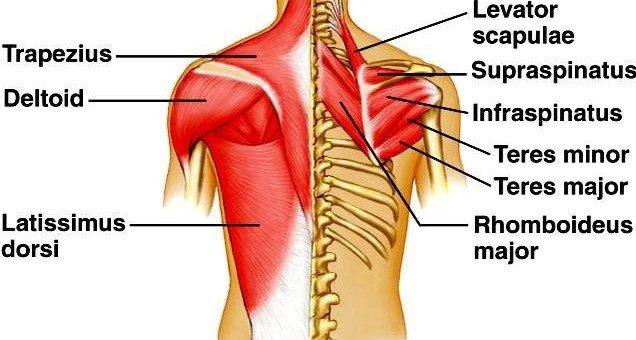
Muscles Move And Support The Spine from www.spineuniverse.com Some of the links in the post above are affiliate links.. As you can see, there are also have a spine of scapula deltoid, triceps brachii, latissimus dorsi. Deep back muscles superficial back muscles action movements of the shoulder. The following diagram below is the diagrams of back muscle. Deep back muscles diagram the superficial layer contains the splenius cervicis and splenius capitis muscles. They extend and rotate the head and neck. The part of the nerve that emerges out of the spine is called the nerve root. Most of the time, back muscle pain is diagnosed then treated with little more than a prescription of rest, painkillers and muscle relaxants.
The deep back muscles, also called intrinsic or true back muscles, consist of four layers of muscles:
See back muscles and low back pain. Deep back muscles superficial back muscles action movements of the shoulder. Another common cause of lower back and hip pain is disc injury. Related posts of back muscles chart muscle anatomy diagram. This is a diagram of the larger and more surface muscles of the low back. The fibres attach to the clavicle, acromion and the scapula spine. Back muscles, like any other muscle in the body, require adequate exercise to maintain strength and tone. Muscles of lower back diagram. Important muscles of the lumbar spine include. Support and protect your spine; Back muscles, back muscle diagram. Chronic back pain map this tool recommended for: The human back extends from the buttocks to the posterior portion of the neck and shoulders.
Superficial, intermediate, deep and deepest layers.these muscles lie on each side of the vertebral column, deep to the thoracolumbar fascia they span the entire length of the vertebral column, extending from the cranium to the pelvis Anatomy muscles view 12 photos of the anatomy muscles view anatomy muscles view, anatomy of body muscles back view, muscle anatomy anterior view, muscle anatomy back view, muscle anatomy posterior view, human muscles, anatomy muscles view, anatomy of body muscles back view, muscle anatomy anterior view. The deep back muscles, also called intrinsic or true back muscles, consist of four layers of muscles: The back muscles enable you to stand up straight; This is a diagram of the larger and more surface muscles of the low back.
Back Muscle Anatomy Science Online from www.online-sciences.com Pain log more pain mapping tools The back consists of the spine, spinal cord, muscles, ligaments, and nerves. Some of the links in the post above are affiliate links.. Daniel nelson on january 1, 2019 2 comments 🔥! Muscle strain is often the cause of back pain from heavy lifting or vigorous exercise. Chronic back pain map this tool recommended for: Intermediate back muscles and c. The back muscles enable you to stand up straight;
Below you'll see diagrams along with the names of the back muscles that may be the cause of your pain.
There are several different layers of muscles in your back that are often pulling in different and various directions. While muscles like the gluteals (in the thighs) are used any time we walk or climb a step, deep back muscles and abdominal muscles are usually not actively engaged during everyday activity. Muscle strain is often the cause of back pain from heavy lifting or vigorous exercise. The part of the nerve that emerges out of the spine is called the nerve root. The back anatomy includes the latissimus dorsi, trapezius, erector spinae, rhomboid, and the teres major. Others, like sumo deadlifts, have been shown in emg studies—and in the trenches—to focus more on other muscle groups than the back. The back comprises the dorsal part of the neck and the torso (dorsal body cavity) from the occipital bone to the top of the tailbone. The deep back muscles, also called intrinsic or true back muscles, consist of four layers of muscles: It joins the lower limb to the pelvic girdle. The pelvis at the bottom of the back and the shoulders at the top of the back give the back. To learn more about the anatomy of the spine, watch this video. Deep back muscles diagram the superficial layer contains the splenius cervicis and splenius capitis muscles. Nerves in your lower back.
They extend and rotate the head and neck. Intermediate back muscles and c. The fibres attach to the clavicle, acromion and the scapula spine. We hope this picture anatomy of back muscles diagram can help you study and research. The deep back muscles, also called intrinsic or true back muscles, consist of four layers of muscles:
Muscles Of The Shoulder And Back Laminated Anatomy Chart Amazon Com Industrial Scientific from images-na.ssl-images-amazon.com Most of the time, back muscle pain is diagnosed then treated with little more than a prescription of rest, painkillers and muscle relaxants. See back muscles and low back pain. This is a table of skeletal muscles of the human anatomy. For more anatomy content please follow us and visit our website: The back comprises the dorsal part of the neck and the torso (dorsal body cavity) from the occipital bone to the top of the tailbone. The back consists of the spine, spinal cord, muscles, ligaments, and nerves. The human back extends from the buttocks to the posterior portion of the neck and shoulders. Anatomynote.com found anatomy of back muscles diagram from plenty of anatomical pictures on the internet.
These structures work together to support the body, enable a range of movements, and send messages from the.
The latissimus dorsi, also known as the lats or wings, are. Pain log more pain mapping tools Both the deltoid and the trapezius are firmly attached to the spine of the scapula. Back of the head muscle structure and nerve system diagram. Muscle strain is often the cause of back pain from heavy lifting or vigorous exercise. Lower back muscle diagram anatomy does degenerative disc disease affect the lower back muscle? In this image, you will find an occipital bone, sternocleidomastoid, trapezius, deltoid in muscles of the lower back diagram. The muscles of the back that work together to support the spine, help keep the body upright and allow twist and bend in many directions. The muscles of the lower back help stabilize, rotate, flex, and extend the spinal column, which is a bony tower of 24 vertebrae that gives the body structure and houses the spinal cord. This is a diagram of the larger and more surface muscles of the low back. Back to tracking tools main page. The intermediate layer contains the erector spinae muscles, whose many functions include the extension and lateral flexion of the spine, head and neck. Diagram lower back wiring diagram data val.
Share :
Post a Comment
for "Back Muscle Diagram / Pin On Health"


Post a Comment for "Back Muscle Diagram / Pin On Health"